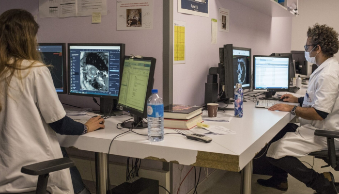
How Radiologists Are Testing For Covid 19 And The Benefits Of Radiology Transcription Software
With 2019-nCoV Pneumonia on the rise globally, researchers have found ways to predict this virus through computed tomography (CT) evidence. A study showed CT imaging was able to identify coronavirus before patients were tested positive. The detection of novel coronavirus has been done with the help of reverse transcription-polymerase chain reaction (RT-PCR). This technique involves the reverse transcription of RNA into DNA and amplification of specific DNA targets to identify DNA that matches the virus. The objective of this study was to describe CT imaging features of five patients who tested negative for coronavirus in RT-PCR tests, or had weak positive results, but were highly suspected of infection. The study observed 167 patients; of these 4 percent had tested negative RT-PCR, but had positive chest CT with a pattern synonymous with viral pneumonia. After positive CT results, all patients were isolated for 2019-nCoV pneumonia. However, in 93 percent of patients, both RT-PCR and CT were positive for 2019-nCoV infection. The authors commented that while a swab test is the standard testing for diagnosis, current tests are majorly time-consuming and facing paucity due to high demand. Additionally, the RT-PCR test for coronavirus may show false negative due to laboratory error or insufficient viral material in the sample.
The study concluded: Previously recorded radiographic studies had shown that most of the cases had analogous features on CT images, like ground-glass opacities (GGO) or mixed GGO. 2019-nCov pneumonia is likely to have a peripheral distribution with bilateral, multifocal lower lung involvement. In the context of usual clinical presentation and exposure to other individuals with 2019-nCoV, CT features of viral pneumonia may be strongly suspicious for 2019-nCoV infection despite negative RT-PCR results. In such cases, repeating swab testing and isolating the patient must be considered.
Radiology published another study regarding the same, “Time Course of Lung Changes on Chest CT During Recovery from 2019 Novel Coronavirus (COVID-19) Pneumonia,” on Feb. 13, 2020. The purpose here was to determine the change in chest CT findings associated with COVID-19 pneumonia from initial diagnosis until patient recovery. Patients with COVID-19 were observed at roughly 4-day intervals from the disease onset till recovery. Patients with pneumonia complications including severe respiratory distress (respiratory rate >30 breaths/min), the requirement for oxygen treatment or mechanical ventilation, or SpO2 lower than 90 percent on room air were excluded. In patients without the severe respiratory disease, the major pulmonary CT findings of novel coronavirus pneumonia were GGO, crazy-paving pattern, and consolidation predominantly in subpleural locations in the lower lobes. Chest CT abnormalities showed an increase in frequency and intensity of lesions in the first 10 weeks (peaking at approximately 10 days). Eventually, there was a short plateauing phase and a decrease in aberrations
How Augnito can help you
Recently, speech recognition (SR) solutions have advanced so much that they are now a practical method of creating radiology reports. More and more departments are now using this technology, and it is rapidly gaining popularity.
Creating reports with radiology transcription software
Using radiology transcription software, the radiologist can dictate the case, edit, and make the final report ready immediately. Hence, the clinician can review the report sooner than it would have been possible had he been using the traditional reporting method. This leads to better patient care since the patient can move on to the next step or start treatment earlier. It can also lead to a more accurate report because it is completed while the radiologist is still reviewing the images in front of him before the details of the case might get forgotten. Additionally, there is a lesser chance of the report getting lost within the system. Finally, this process can lead to a more satisfied referring physician, since the report is available immediately as well as a more content radiologist, as the report will not have to be reviewed again.
Benefits of radiology transcription software
The two main benefits are faster completion of reports, and reduced number of transcriptionists needed. Another advantage is the lack of need for calling regarding preliminary reports. Additionally, radiology staff spends less time looking for film, since the referring physician will have the report almost immediately. Finally, it allows the radiologist more control over the dictation process since they don’t rely on a transcriptionist and also don’t need to create an initial “verbal” report before completing a “final” report eventually.
Rapid report creation
Numerous studies have been conducted to ascertain how much faster reports are completed using radiology transcription software. One study stated that the mean report turnaround time improved from 87.8 to 43.6 hours. The study concluded that report availability at 24 hours increased from 10.5% to 62.5%. In another study, report creation time was reduced from roughly 2 to 4 hours with transcriptionists to less than 5 minutes with the aid of radiology transcription software.
Another study investigated the use of these systems in a teaching hospital, where cases are first dictated by residents and then passed to the staff radiologist for final acceptance. The researchers noted that when attending physicians dictated studies themselves, 65% of reports were completed within 15 minutes and 90% of the time in less than an hour. Using traditional transcriptionists, the mean report turnaround time was 30 hours. However, studies showed that the final report was available in less than an hour only 15% of the time and in 90% of cases, reports were available within 5 hours. The delay between the time that the resident completed the report and the time that the attending radiologist signed off on it was significant.
Free transcriptionists
Radiology transcription software reduces the number of transcriptionists used by the department. This fact has been noted by two studies that concluded that departments almost always save money after implementing these systems. The first study states savings of $100,000. The second study observed that their department saved $1.7 million within the first 5 years.
Conclusion
Radiology transcription software is now a feasible option for most users, in terms of both precision and user-friendliness. The advantages of better report turnaround time and cost-effectiveness must be measured against the increased time radiologists spend editing reports.

